
Viruses
[Page content sourced from CDC]
Drug-Resistant Organisms
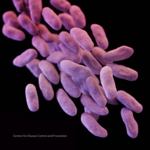
Acinetobacter
Campylobacter

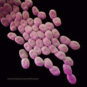
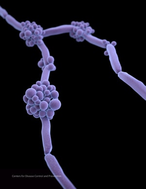
Candida
Carbapenem-resistant Enterobacteriaceae
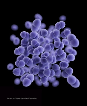
Enterococcus
Extended-spectrum β-lactamase
Tuberculosis

MRSA

PHIL ID # 10046
Photo Credit: Janice Haney, Centers for Disease Control and Prevention
Download High Resolution
Description:
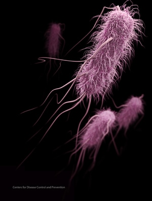
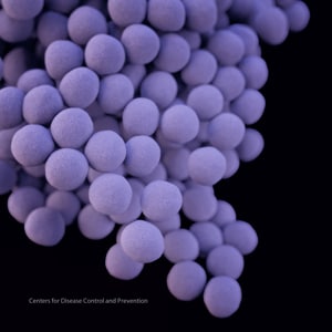
This 2005 colorized scanning electron micrograph (SEM) depicts numerous clumps of methicillin-resistant Staphylococcus aureus (MRSA) bacteria. Methicillin-resistant Staphylococcus aureus infections, e.g., bloodstream, pneumonia, bone infections, occur most frequently among persons in hospitals and healthcare facilities, including nursing homes, and dialysis centers.
MRSA
PHIL ID # 10045
Photo Credit: Janice Carr, Centers for Disease Control and Prevention
Download High Resolution
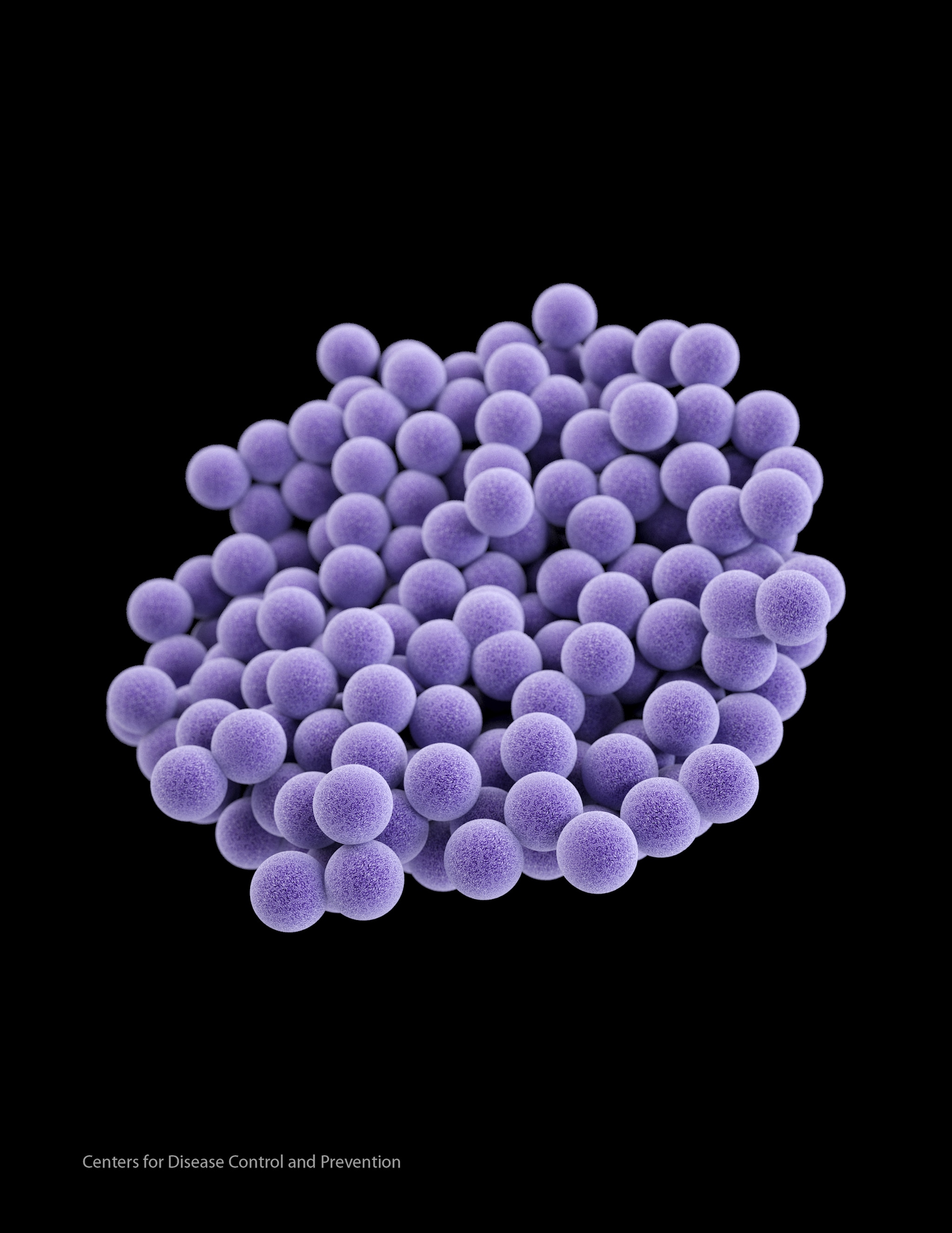
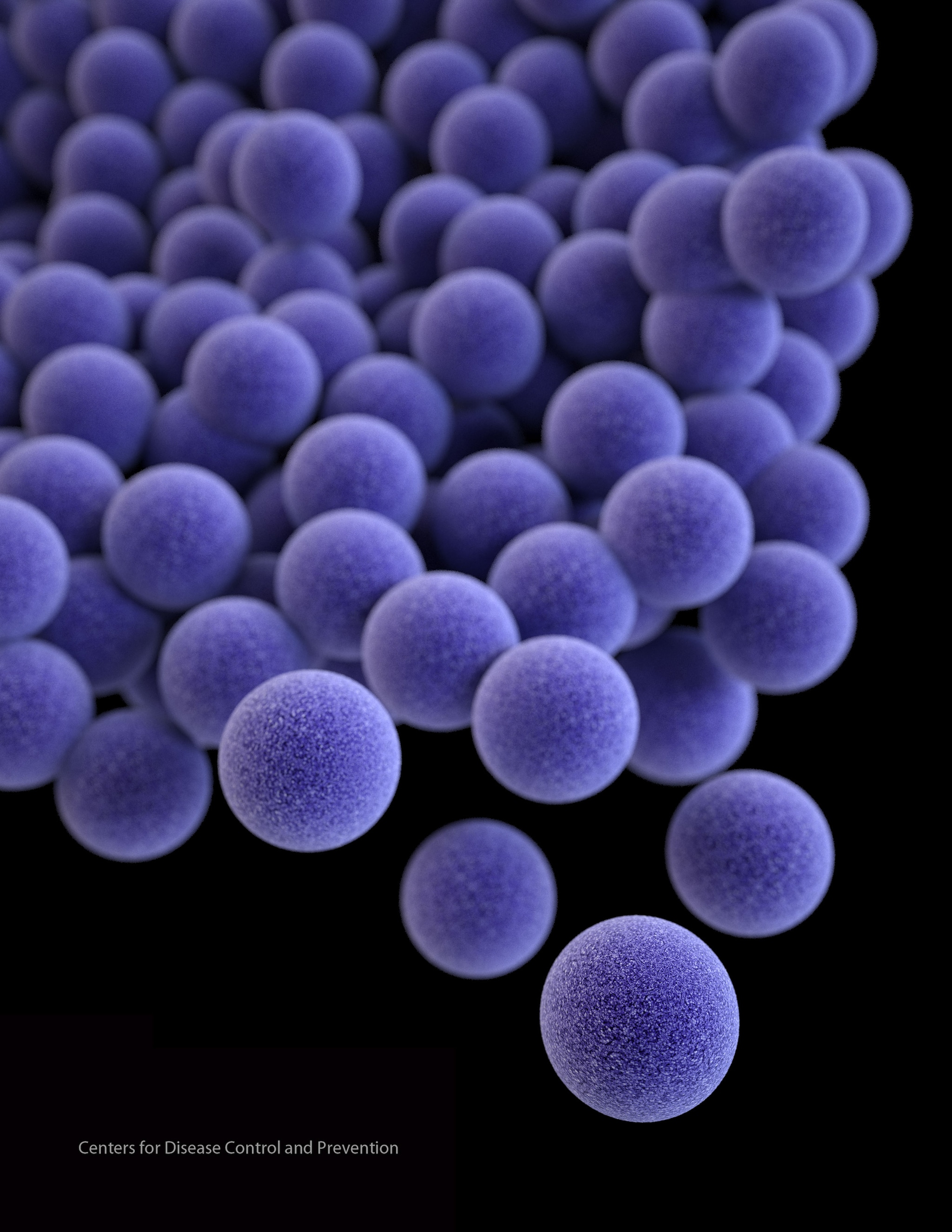
Description: This 2005 colorized scanning electron micrograph (SEM) depicts numerous clumps of methicillin-resistant Staphylococcus aureus (MRSA) bacteria. Methicillin-resistant Staphylococcus aureus infections, e.g., bloodstream, pneumonia, bone infections, occur most frequently among persons in hospitals and healthcare facilities, including nursing homes, and dialysis centers.


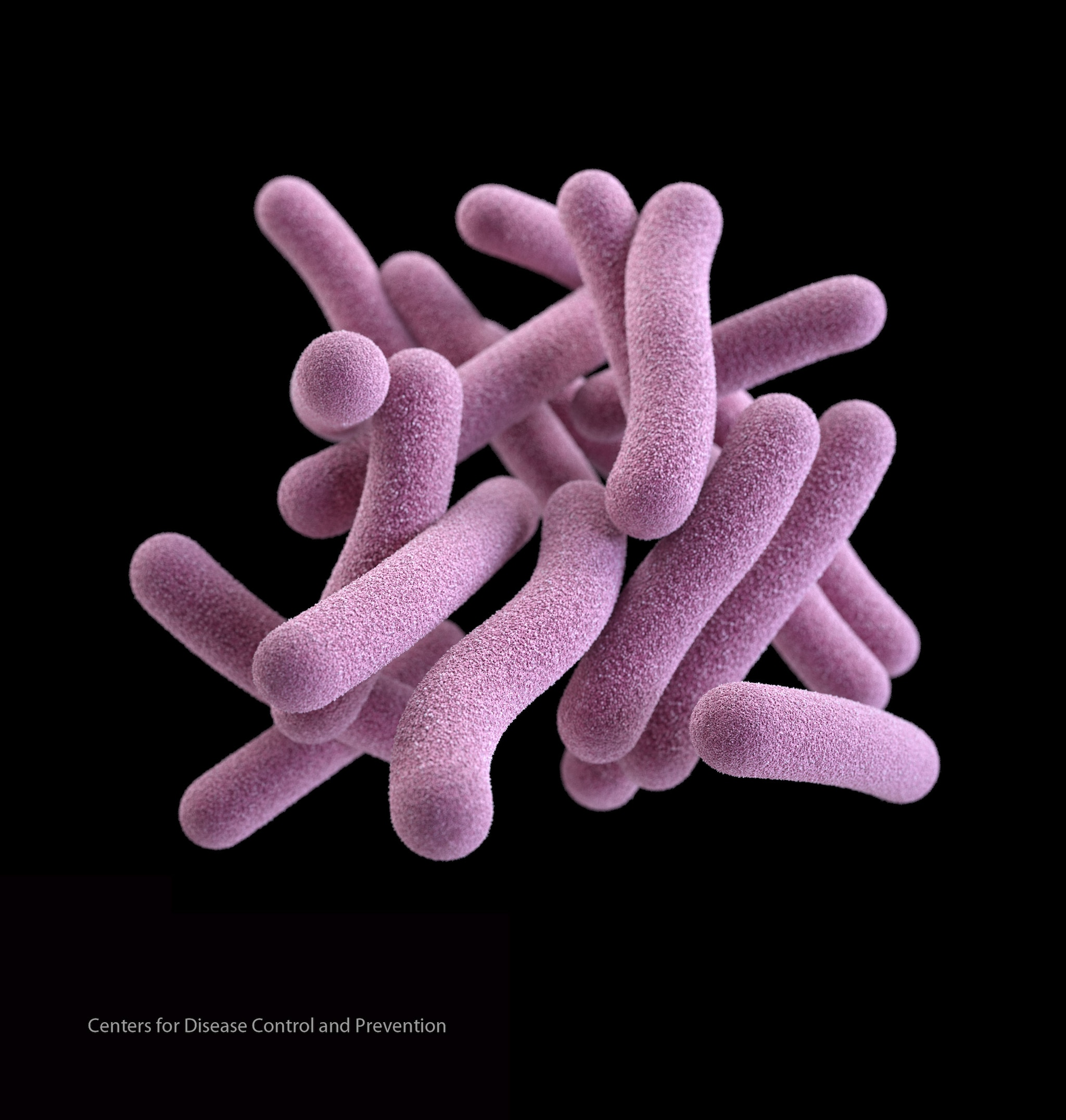
Tuberculosis
Common and Recent Organisms
Zika Virus


PHIL ID #20487
Photo Credit: Cynthia Goldsmith
Download High Resolution
Description:
This is a transmission electron micrograph (TEM) of Zika virus, which is a member of the family Flaviviridae. Virus particles are 40 nm in diameter, with an outer envelope, and an inner dense core.
Ebola

PHIL ID #10815
Photo Credit: Frederick Murphy
Download High Resolution
Description:
Colorized transmission electron micrograph (TEM) revealing some of the ultrastructural morphology displayed by an Ebola virus virion.
Ebola

PHIL ID #10816
Photo Credit: Frederick Murphy
Download High Resolution
Description:
Colorized transmission electron micrograph (TEM) revealing some of the ultrastructural morphology displayed by an Ebola virus virion.
Avian Influenza A H7N9

PHIL ID #15670
Photo Credit: Cynthia S. Goldsmith and Thomas Rowe
Download High Resolution
Description:
Influenza A H7N9 as viewed through an electron microscope. Both filaments and spheres are observed in this photo.
2009 H1N1 Flu

PHIL ID #11822
Photo Credit: Illustrator: Dan Higgins, CDC
Download High Resolution
Description:
This picture provides a 3D graphic representation of a generic influenza virion’s ultrastructure and is not specific to a seasonal, avian, or 2009 H1N1 virus. Note the key to the right identifying the virion’s surface protein constituents. See PHIL 11823 for an uncut view of the virion’s exterior.
2009 H1N1 Flu

PHIL ID #11823
Photo Credit: Illustrator: Dan Higgins, CDC
Download High Resolution
Description:
This picture provides a 3D graphic representation of a generic influenza virion’s ultrastructure and is not specific to a seasonal, avian, or 2009 H1N1 virus. See PHIL 11822 for a view of this virus in which a portion of the virion’s protein coat, or “capsid”, has been cut away, revealing its inner nucleic acid core proteins, as well as a key identifying the organism’s protein constituents.
2009 H1N1 Flu

PHIL ID #11215
Photo Credit: C. S. Goldsmith and A. Balish, CDC
Download High Resolution
Description:
Negative stain EM image of the 2009 H1N1 influenza A/CA/4/09
2009 H1N1 Flu

PHIL ID #11214
Photo Credit: C. S. Goldsmith and A. Balish, CDC
Download High Resolution
Description:
Negative stain EM image of the 2009 H1N1 Influenza A/CA/4/09
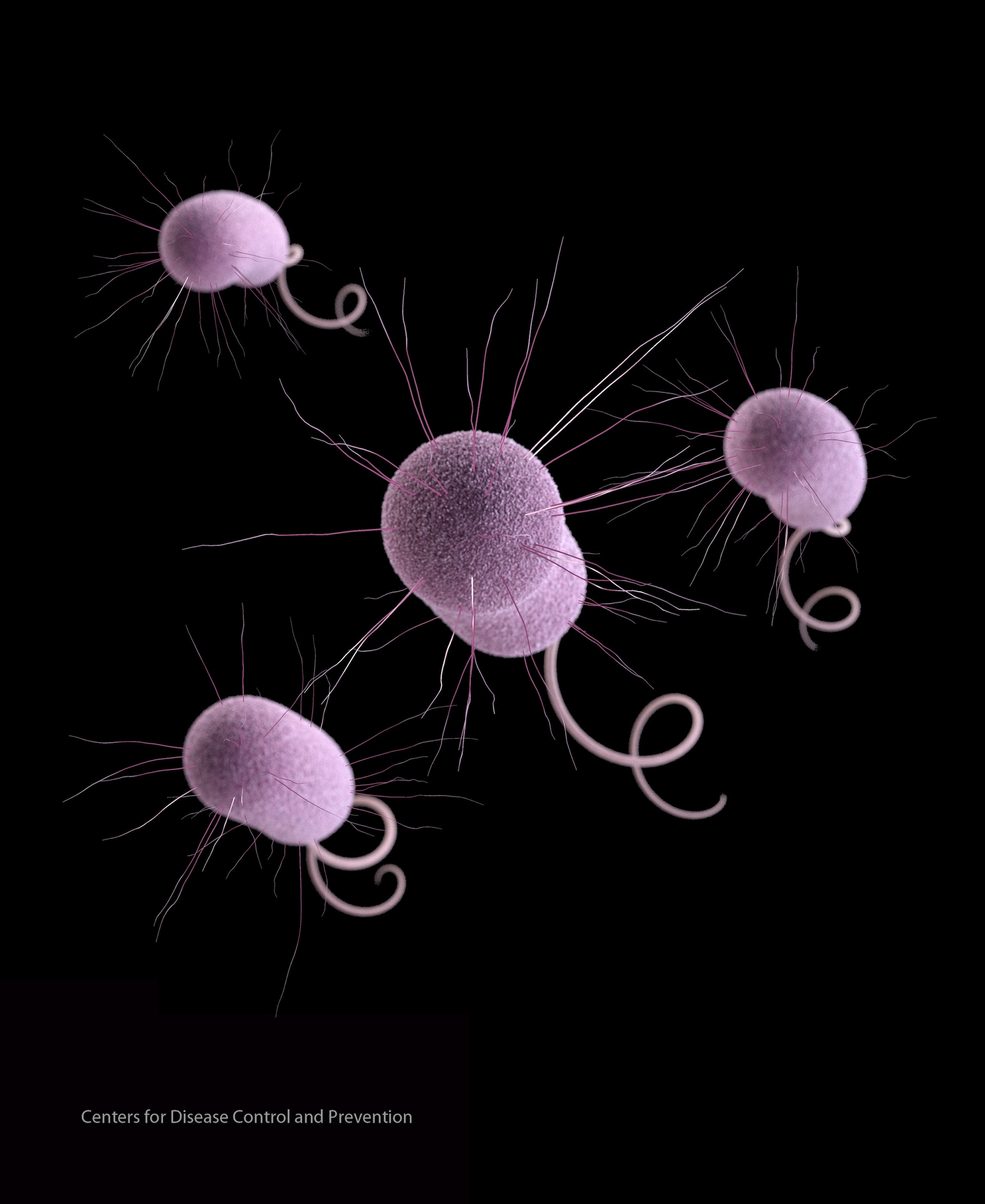
Legionella (Legionnaire′s Disease)
 PHIL ID #11152
Photo Credit: Janice Haney Carr, Centers for Disease Control and Prevention
Download High Resolution
Description:
This image depicts a large grouping of Gram-negative Legionella pneumophila bacteria.
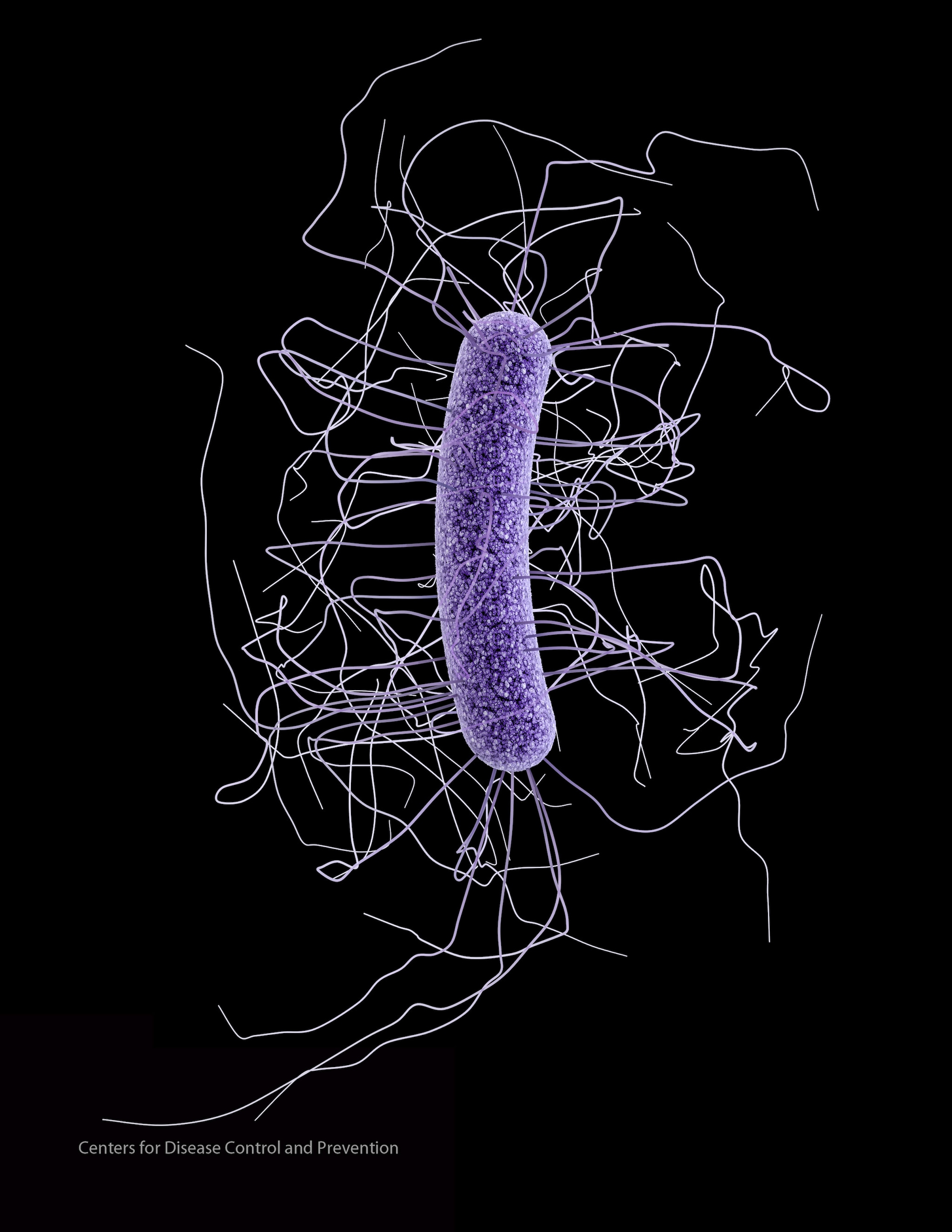
Clostridium difficile
 PHIL ID #9999
Photo Credit: Janice Haney Carr, Centers for Disease Control and Prevention
Download High Resolution
Description: This micrograph depicts gram-positive Clostridium difficile bacteria from a stool sample culture obtained using a .1µm filter.
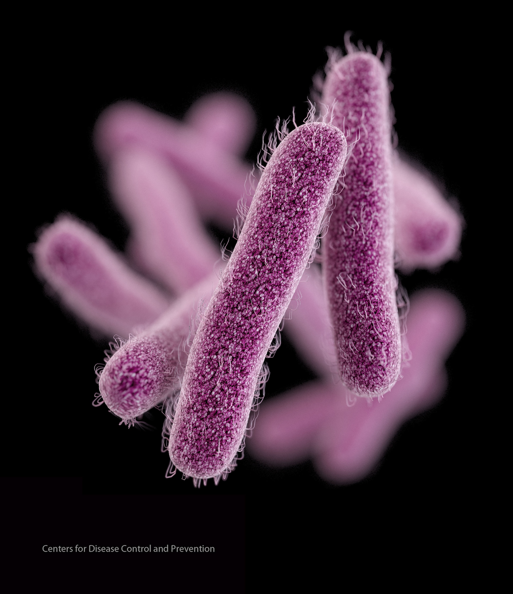
Salmonella Typhimurium

PHIL ID #10983
Photo Credit: Janice Haney Carr, Centers for Disease Control and Prevention
Download High Resolution
Description:
This colorized scanning electron micrograph (SEM) depicts a colony of Salmonella Typhimurium bacteria.
Salmonella Typhimurium

PHIL ID #10971
Photo Credit: Janice Haney Carr, Centers for Disease Control and Prevention
Download High Resolution
Description:
This colorized scanning electron micrograph (SEM) depicts a colony of Salmonella Typhimurium bacteria.
Salmonella

PHIL ID #10896
Photo Credit: Janice Haney Carr, Centers for Disease Control and Prevention
Download High Resolution
Description:
This colorized scanning electron micrograph (SEM) depicts a colony of rod-shaped Salmonella sp. bacteria
Influenza virus particle

PHIL ID #10073
Photo Credit: Cynthia Goldsmith, Centers for Disease Control and Prevention
Download High Resolution
Description:
This negative-stained transmission electron micrograph (TEM) depicts the ultrastructural details of an influenza virus particle, or “virion.”
West Nile Virus

PHIL ID #10700
Photo Credit: Cynthia Goldsmith, Centers for Disease Control and Prevention
Download High Resolution
Description:
This transmission electron micrograph (TEM) depicts the presence of West Nile virus virions.
West Nile Virus

PHIL ID #10701
Photo Credit: Cynthia Goldsmith, Centers for Disease Control and Prevention
Download High Resolution
Description:
This is a transmission electron micrograph (TEM) of the West Nile virus (WNV). See PHIL 2290 for a black and white version of this image.
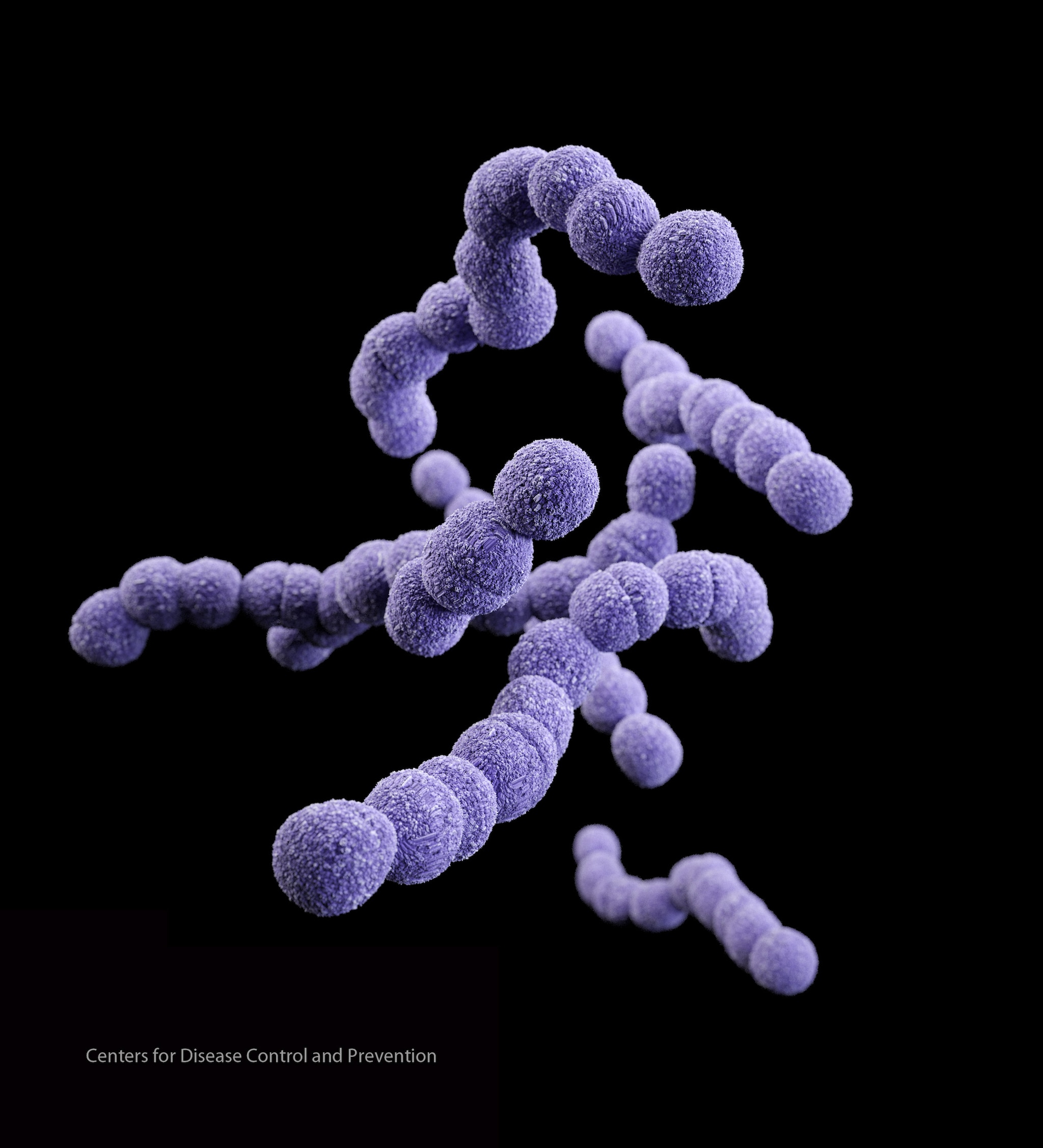
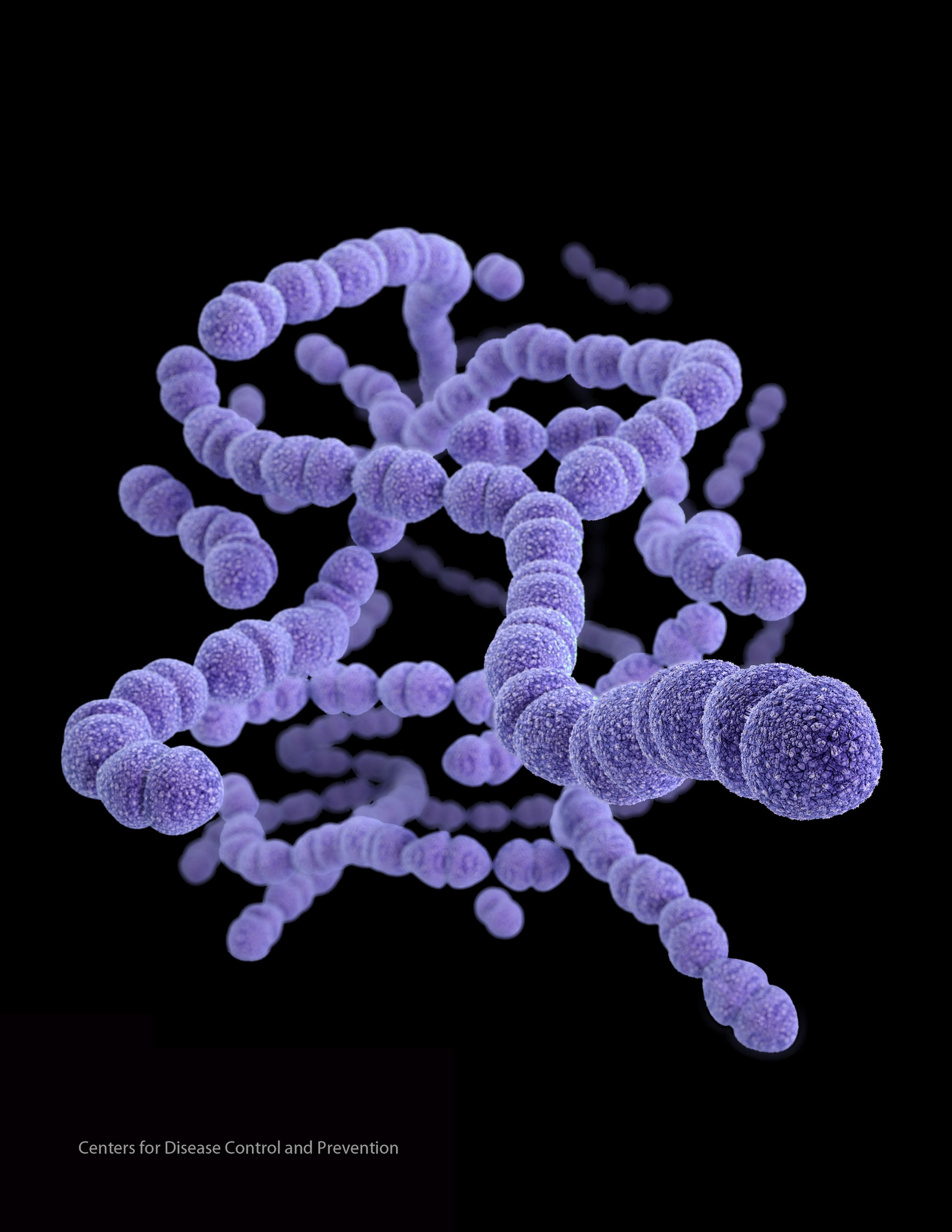
Group C Streptococcus

PHIL ID #10586
Photo Credit: Janice Haney Carr, Centers for Disease Control and Prevention
Download High Resolution
Description:
This colorized scanning electron micrograph (SEM) reveals a small clustered group of gram-positive, beta-hemolytic Group C Streptococcus sp. bacteria. See PHIL 10585 for a black and white version of this image.
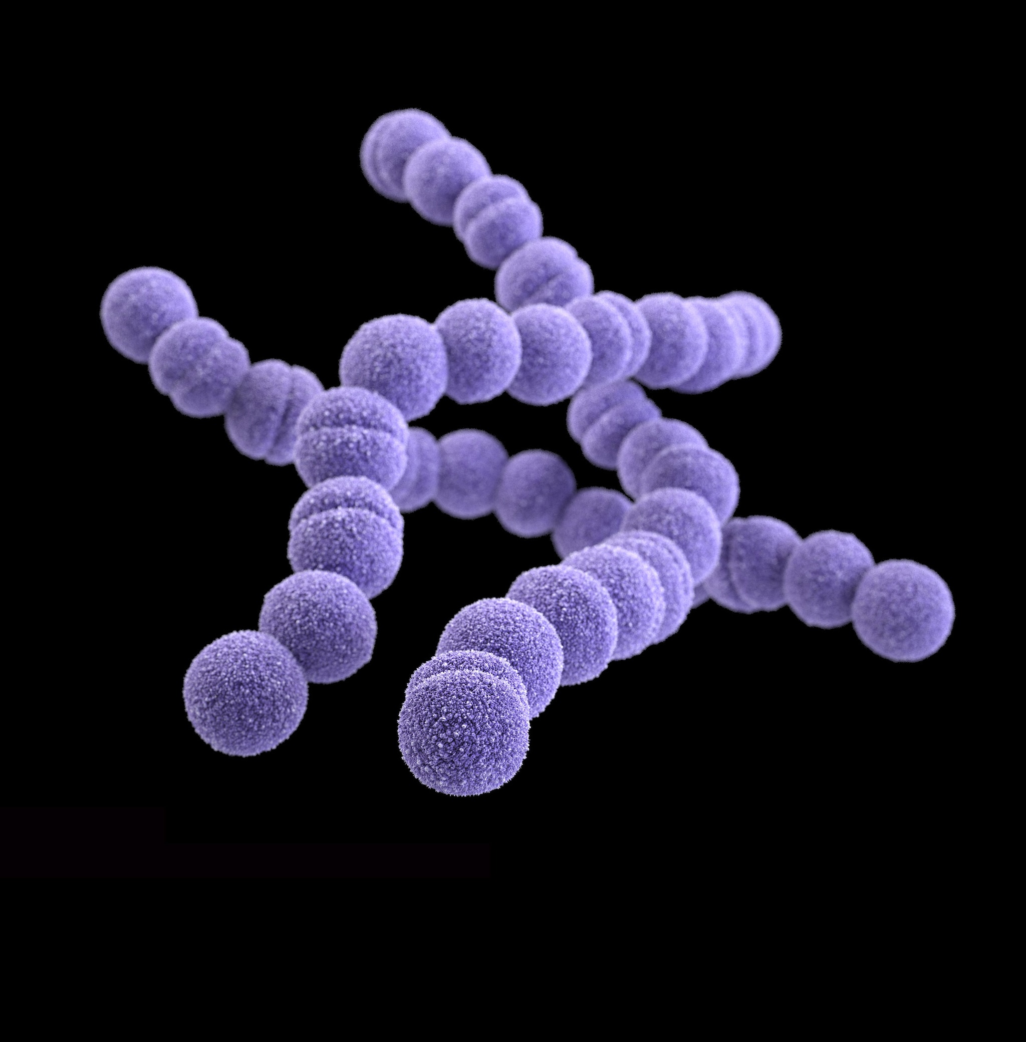
Group C Streptococcus

PHIL ID #10591
Photo Credit: Janice Haney Carr, Centers for Disease Control and Prevention
Download High Resolution
Description:
This colorized scanning electron micrograph (SEM) reveals a small clustered group of Gram-positive, beta-hemolytic Group C Streptococcus sp. bacteria. See PHIL 10585 for a black and white version of this image
Measles

PHIL ID #10707
Photo Credit: Cynthia Goldsmith, Centers for Disease Control and Prevention
Download High Resolution
Description:
This thin-section transmission electron micrograph (TEM) reveals a single virus particle, or virion, of measles virus.
Norovirus

PHIL ID #10708
Photo Credit: Charles D. Humphrey, Centers for Disease Control and Prevention
Download High Resolution
Description:
This transmission electron micrograph (TEM) reveals norovirus virions, or virus particles.
Norovirus

PHIL ID #10709
Photo Credit: Charles D. Humphrey, Centers for Disease Control and Prevention
Download High Resolution
Description:
This transmission electron micrograph (TEM) reveals norovirus virions, or virus particles.
Avian Influenza A H5N1

PHIL ID # 1841
Photo Credit: Cynthia Goldsmith, Centers for Disease Control and Prevention
Download High Resolution
Description:
Colorized transmission electron micrograph of avian influenza A H5N1 viruses (seen in gold) grown in MDCK cells (seen in green).
Avian influenza A viruses do not usually infect humans; however, several instances of human infections and outbreaks have been reported since 1997. When such infections occur, public health authorities monitor these situations closely.
E. Coli

PHIL ID # 10068
Photo Credit: Janice Haney Carr, Centers for Disease Control and Prevention
Download High Resolution
Description:
This colorized scanning electron micrograph (SEM) depicts a number of Escherichia coli bacteria of the strain O157:H7. This strain of E. coli is an emerging cause of foodborne illness. An estimated 73,000 cases of infection, and 61 deaths occur in the United States each year. Infection often leads to bloody diarrhea, and occasionally to kidney failure. Most illness has been associated with eating undercooked, contaminated ground beef. Person-to-person contact in families and child care centers is also an important mode of transmission. Infection can also occur after drinking raw milk, and after swimming in, or drinking sewage-contaminated water.
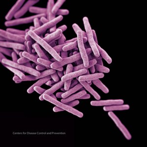
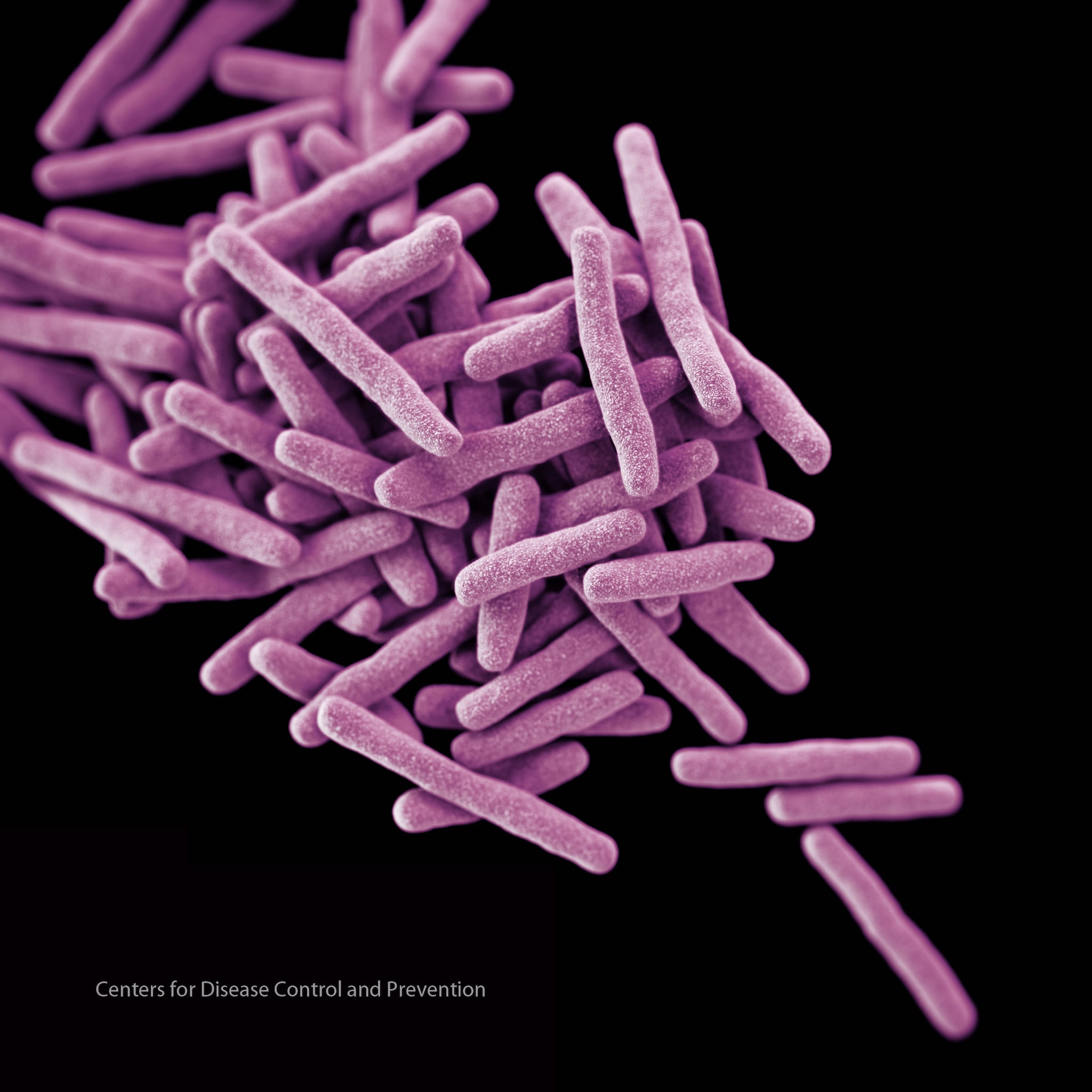
Tuberculosis

PHIL ID # 9997
Photo Credit: Janice Carr, Centers for Disease Control and Prevention
Download High Resolution
Description:
This colorized scanning electron micrograph (SEM) depicted some of the ultrastructural details seen in the cell wall configuration of a number of Gram-positive Mycobacterium tuberculosis bacteria. TB bacteria become active, and begin to multiply, if the immune system can’t stop them from growing. The bacteria attack the body and destroy tissue. If in the lungs, the bacteria can actually create a hole in the lung tissue. Some people develop active TB disease soon after becoming infected, before their immune system can fight off the bacteria. Other people may get sick later, when their immune system becomes weak for another reason.
HIV-1

PHIL ID # 10000
Photo Credit: Cynthia Goldsmith, Centers for Disease Control and Prevention
Download High Resolution
Description:
Scanning electron micrograph of HIV-1 budding from cultured lymphocyte. See PHIL 1197 for a black and white view of this image.

KB said
Worldwide emergence of resistance to antifungal drugs challenges human health and food security
Matthew C. Fisher et al.
Science 18 May 2018:
Vol. 360, Issue 6390, pp. 739-742
DOI: 10.1126/science.aap7999
http://science.sciencemag.org/content/360/6390/739.full
Abstract
The recent rate of emergence of pathogenic fungi that are resistant to the limited number of commonly used antifungal agents is unprecedented. The azoles, for example, are used not only for human and animal health care and crop protection but also in antifouling coatings and timber preservation. The ubiquity and multiple uses of azoles have hastened the independent evolution of resistance in many environments. One consequence is an increasing risk in human health care from naturally occurring opportunistic fungal pathogens that have acquired resistance to this broad class of chemicals. To avoid a global collapse in our ability to control fungal infections and to avoid critical failures in medicine and food security, we must improve our stewardship of extant chemicals, promote new antifungal discovery, and leverage emerging technologies for alternative solutions.
The rapid emergence of multidrug-resistant pathogenic fungi and the better-publicized threat of antibiotic-resistant bacteria together pose a considerable threat to disease control across diverse anthropogenic systems. These microbes respond adroitly to human-induced natural selection through chemical treatments and nimbly hijack human globalization pathways (1), thus disseminating the problems worldwide. Today, crop-destroying fungi account for perennial yield losses of ~20% worldwide, with a further 10% loss postharvest. Fungal effects on human health are currently spiraling, and the global mortality rate for fungal diseases now exceeds that for malaria or breast cancer and is comparable to those for tuberculosis and HIV (2). Fungal infections have hitherto been greatly neglected relative to other classes of infectious disease, despite their ubiquity.
http://science.sciencemag.org/content/360/6390/739.full
te2ataria said
The deadly brainwashing power of parasites
“These parasites are highly specialised and have evolved a fascinating array of approaches to manipulate their hosts.”
http://www.nzherald.co.nz/nz/news/article.cfm?c_id=1&objectid=11744312